Digital Histology Biology Diagrams Additionally, localized protein synthesis to support spindle formation is achieved in the spindle forming region, whilst protein synthesis is reduced elsewhere in the ooplasm. This is achieved through enrichment of spindle-related mRNAs in the spindle forming region combined with local PLK1-mediated phosphorylation and inactivation of the
Centromere-telomere interchangeability in promoting meiotic spindle formation. Meiotic progression is represented from left to right. (A) In WT meiocytes, the movement of telomeres to the SPB promotes modification of Sad1 (yellow stars) or the NM surrounding Sad1, in turn promoting Sad1 accumulation and ensuring bipolar spindle formation.
Oocyte Meiotic Spindle Assembly and Function Biology Diagrams
The universal features of meiotic spindle formation have yet to be determined; nonetheless, modulation of this process by the bouquet may occur in many eukaryotes. In higher eukaryotes, meiotic spindles can be nucleated from several different structures, including centrosomes, MTOCs, and complexes of motor proteins. Expressed throughout meiotic progression, PLK1 promotes meiotic progression and is needed for high-quality meiotic spindle formation (Pahlavan et al., 2000; Solc et al., 2015). Overall, the important role of PLK1 in oocyte meiosis, its responsiveness to exogenous and endogenous signals, and the known adverse effects of these signals on meiotic Meiotic spindle abnormality in human oocytes is a hallmark of maternal ageing [44], [196]. In conclusion, it is clear that there are gene products that are essential for control of spindle formation, chromosome cohesion and chromosome segregation in meiosis. These possibly present targets for chemicals that specifically interfere with
Herein, we describe the recent advances in understanding the mechanisms controlling formation of the meiotic spindle in metaphase I (MI) and metaphase II (MII) in mammalian oocytes, and focus on the differences between mouse and human oocytes. Unlike mitotic cells, mammalian oocytes lack typical centrosomes that consist of two centrioles and

PDF Oocyte Meiotic Spindle Assembly and Function Biology Diagrams
Although centrosome-mediated spindle formation is dominant in most mitotic cells, mitosis can still take place in the absence of centrosomes, showing that other centrosome-independent pathways can participate in spindle formation (Khodjakov et al., 2000; Basto et al., 2006; Azimzadeh et al., 2012; Bazzi and Anderson, 2014). These centrosome
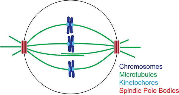
Kinetochore fibers are not involved in the formation of the first meiotic spindle in mouse oocytes, but control the exit from the first meiotic M phase. The Journal of Cell Biology, 146, 1-12. [PMC free article] [Google Scholar] Burbank KS, et al. (2006). A new method reveals microtubule minus ends throughout the meiotic spindle.